Microangiopathic hemolytic anemia is a rare blood disorder. It happens when red blood cells get destroyed and small blood clots form in tiny blood vessels. This can greatly affect a person’s health and needs careful medical attention. We will look into what this condition is, its types, how it happens, how to diagnose it, and how to treat it.
Microangiopathic hemolytic anemia is a rare blood disorder characterized by the destruction of red blood cells and the formation of small blood clots.
The condition can have various underlying causes, including thrombotic microangiopathies, hemolytic uremic syndrome, and thrombotic thrombocytopenic purpura.
Endothelial injury and red blood cell fragmentation play a crucial role in the pathogenesis of microangiopathic hemolytic anemia.
Diagnosis involves a comprehensive evaluation of laboratory tests, imaging studies, and clinical manifestations.
Management strategies include supportive care, targeted therapies, and addressing the underlying cause of the condition.
Introduction to Microangiopathic Hemolytic Anemia
Microangiopathic hemolytic anemia is a serious blood disorder. It happens when red blood cells break apart in small blood vessels. This can cause serious problems, like organ damage and failure.
Definition and Significance
This type of anemia means red blood cells get destroyed as they go through small blood vessels. This is called intravascular hemolysis. It’s important because it can block blood flow and oxygen to important organs. This leads to serious health issues.
Types of Microangiopathic Hemolytic Anemia
There are different types of microangiopathic hemolytic anemia. Thrombotic microangiopathies are one kind. They happen when small blood clots form in tiny blood vessels. This makes red blood cells break apart and can harm organs.
Type of Microangiopathic Hemolytic Anemia
Key Characteristics
Thrombotic Microangiopathies
Characterized by the formation of small blood clots in the small blood vessels, leading to red blood cell fragmentation and organ damage.
Hemolytic Uremic Syndrome
A specific type of thrombotic microangiopathy that primarily affects the kidneys, often caused by Shiga toxin-producing bacteria.
Thrombotic Thrombocytopenic Purpura
A rare and life-threatening thrombotic microangiopathy characterized by a severe deficiency of the ADAMTS13 enzyme, which leads to the formation of blood clots.
Knowing about the different types of microangiopathic hemolytic anemia helps doctors treat patients better.
Thrombotic Microangiopathies
Thrombotic microangiopathies (TMAs) are serious disorders. They cause small blood clots in tiny blood vessels. This can destroy red blood cells and lead to serious problems. TMAs are key to understanding microangiopathic hemolytic anemia.
TMAs form tiny blood clots that block blood flow to organs. This can damage organs, harm the brain, and even cause failure of multiple organs. Knowing about TMAs helps doctors treat microangiopathic hemolytic anemia.
Types of Thrombotic Microangiopathies
Hemolytic uremic syndrome (HUS)
Thrombotic thrombocytopenic purpura (TTP)
Atypical hemolytic uremic syndrome (aHUS)
Complement-mediated TMA
Pregnancy-associated TMA
Drug-induced TMA
Each TMA type has its own causes and symptoms. Knowing the type helps doctors treat it better. This improves how well patients do.
Type of TMA
Causes
Distinguishing Features
Hemolytic Uremic Syndrome (HUS)
Shiga toxin-producing E. coli, complement dysregulation
Severe thrombocytopenia, neurological manifestations, fever
Atypical Hemolytic Uremic Syndrome (aHUS)
Complement dysregulation
Acute kidney injury, extrarenal organ involvement, recurrent episodes
Knowing the differences between TMA types helps doctors treat patients with microangiopathic hemolytic anemia better.
Hemolytic Uremic Syndrome
Hemolytic uremic syndrome (HUS) is a serious condition caused by the Shiga toxin from some bacteria. It’s marked by anemia, low platelet count, and kidney damage. This makes it a severe health threat.
Causes and Pathophysiology
The main cause of HUS is the Shiga toxin from E. coli O157:H7 bacteria. This toxin harms the blood vessel walls. It causes platelets to activate and form blood clots.
This leads to blockages in small blood vessels. It also causes anemia, low platelet count, and kidney problems.
Some cases can get worse quickly, leading to serious issues like kidney failure and brain problems.
“Prompt recognition and appropriate management of HUS are crucial, as early intervention can significantly improve patient outcomes.”
Quick diagnosis and treatment are key to handling HUS. Without it, the condition can cause severe and long-term health problems.
Thrombotic Thrombocytopenic Purpura
Thrombotic thrombocytopenic purpura (TTP) is a rare and serious condition. It happens when there’s not enough ADAMTS13 enzyme. This enzyme helps control blood clotting. Without enough, abnormal blood clots form in small blood vessels.
ADAMTS13 Deficiency
ADAMTS13 breaks down large von Willebrand factor (vWF) multimers. These multimers can cause clots if they stick together. In TTP, not having enough ADAMTS13 lets these multimers build up.
This buildup causes clots in tiny blood vessels. It also destroys red blood cells and lowers platelet count. These are the main signs of TTP.
Key Aspects of ADAMTS13 Deficiency in Thrombotic Thrombocytopenic Purpura
ADAMTS13 is a metalloprotease that normally regulates blood clotting by breaking down large von Willebrand factor (vWF) multimers.
In TTP, there is a severe deficiency in ADAMTS13 activity, often due to autoantibodies that inhibit the enzyme’s function.
The lack of ADAMTS13 allows the accumulation of large vWF multimers, leading to the formation of small blood clots in the body’s microvasculature.
This process results in the destruction of red blood cells (hemolytic anemia) and a low platelet count (thrombocytopenia), the hallmark features of TTP.
Understanding ADAMTS13’s role in TTP has been a big step forward. It helps us know how to treat this condition. This knowledge leads to better treatments for patients with TTP.
Role of Shiga Toxin
Shiga toxin is a powerful toxin that plays a key role in some types of microangiopathic hemolytic anemia. This includes hemolytic uremic syndrome (HUS). It is made by certain E. coli bacteria, like the O157:H7 type. This toxin can severely harm the human body.
This toxin damages the cells that line blood vessels. It starts the blood clotting process and forms clots. These clots can block blood flow, causing tissue damage and destroying red blood cells. This is a key sign of microangiopathic hemolytic anemia.
Shiga toxin also harms the kidney’s filtering system. It makes it hard for the kidney to remove waste and extra fluids. This can lead to a severe condition called hemolytic uremic syndrome. This condition is marked by thrombocytopenia, microangiopathic hemolytic anemia, and acute kidney injury.
Knowing how shiga toxin causes some microangiopathic hemolytic anemias is vital for doctors. It helps them diagnose and treat these conditions effectively. This can prevent more harm.
“Shiga toxin is a potent bacterial toxin that can trigger the development of certain types of microangiopathic hemolytic anemia, such as hemolytic uremic syndrome.”
Complement Dysregulation in Microangiopathic Hemolytic Anemia
Microangiopathic hemolytic anemia (MAHA) is a complex condition. It causes the destruction of red blood cells in small blood vessels. Research shows that the complement system’s imbalance plays a big role in some types of MAHA.
The complement system helps fight off foreign invaders by recognizing and eliminating them. In MAHA, an imbalance in the complement system can cause it to activate too much. This leads to damage in the blood vessel walls and the destruction of red blood cells.
Complement Dysregulation in Hemolytic Uremic Syndrome
Hemolytic uremic syndrome (HUS) is a type of MAHA linked to complement dysregulation. Mutations or autoantibodies against key proteins like factor H can upset the balance of the complement system. This imbalance can lead to HUS.
Complement Dysregulation in Atypical Hemolytic Uremic Syndrome
Atypical HUS (aHUS) is a rare form of HUS caused by complement dysregulation. Genetic or acquired issues with complement regulators can cause the complement system to activate uncontrollably. This leads to damage in blood vessel walls and the formation of blood clots.
Understanding how complement dysregulation affects MAHA is crucial for finding new treatments. By fixing the complement imbalance, doctors may be able to stop the damage and help patients recover better.
Complement Regulation in MAHA
Pathophysiological Consequences
Genetic or acquired abnormalities in complement regulators
Uncontrolled activation of the complement cascade, leading to endothelial injury and red blood cell destruction
Disruption of the balance in the complement system, contributing to the development of MAHA
Studying complement dysregulation in MAHA has led to new insights. It shows how to tackle the disorder at its core. This could lead to better treatments and care for patients.
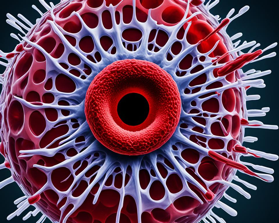
Endothelial Injury and Red Blood Cell Fragmentation
Microangiopathic hemolytic anemia often shows up as red blood cells breaking into pieces, making schistocytes. This happens because the endothelial cells, which line the blood vessels, get hurt and don’t work right.
Mechanisms of Endothelial Damage
The endothelium acts as a barrier between the blood and the body’s tissues. It can get damaged by things like too much pressure, immune reactions, or certain toxins. This damage can start a chain reaction that breaks red blood cells.
Formation of Schistocytes
Breaking of red blood cells, or red blood cell fragmentation, is a key sign of microangiopathic hemolytic anemia. These broken cells, called schistocytes, show up on blood tests and help diagnose the condition. They form because of the first endothelial injury and the stress on red blood cells moving through damaged blood vessels.
Knowing how endothelial injury and red blood cell breaking relate is key to treating microangiopathic hemolytic anemia.
microangiopathic hemolytic anemia
Microangiopathic hemolytic anemia is a complex condition. It happens when red blood cells break apart in small blood vessels. This is called intravascular hemolysis. It’s key to understanding this disorder.
This condition starts with damage to the blood vessel lining. This damage can come from many things like blood clots, immune issues, or certain medicines. When the lining gets damaged, it can’t protect the blood flow well.
This damage leads to tiny blood clots in the small blood vessels. These clots block the flow of red blood cells. When red blood cells try to get past these clots, they break apart. This breaking apart is a sign of microangiopathic hemolytic anemia.
Key Mechanisms in Microangiopathic Hemolytic Anemia
Endothelial injury and dysfunction
Activation of coagulation pathways and formation of microthrombi
Mechanical fragmentation of red blood cells (schistocyte formation)
Intravascular hemolysis and release of hemoglobin
Understanding these mechanisms helps doctors diagnose and treat microangiopathic hemolytic anemia. This can improve how well patients do and their quality of life.
Diagnostic Approach
Diagnosing microangiopathic hemolytic anemia requires a detailed look at both lab tests and imaging studies. These tools help find the cause and plan the right treatment.
Laboratory Tests
The main tests for this condition are:
Complete blood count (CBC) to check anemia and low platelet levels
Peripheral blood smear to spot schistocytes (broken red blood cells)
Lactate dehydrogenase (LDH) levels, often high due to red blood cell destruction
Haptoglobin levels, usually low from the destruction of red blood cells
Kidney function tests, like BUN and creatinine, to check for kidney problems
Imaging Studies
Imaging studies give important clues in diagnosing microangiopathic hemolytic anemia. They include:
Abdominal ultrasound to check for issues in organs like the kidneys or liver
Computed tomography (CT) scans or magnetic resonance imaging (MRI) for conditions like blood clots or organ damage
Echocardiography to check the heart’s function and rule out heart issues
Together, lab tests and imaging studies help doctors make a diagnosis. They find the cause and plan the best treatment for patients with microangiopathic hemolytic anemia.
Test
Purpose
Complete Blood Count (CBC)
Assess anemia and thrombocytopenia
Peripheral Blood Smear
Detect schistocytes (fragmented red blood cells)
Lactate Dehydrogenase (LDH)
Evaluate for increased red blood cell destruction
Haptoglobin
Assess for decreased levels due to hemolysis
Kidney Function Tests (BUN, Creatinine)
Evaluate for renal impairment
Abdominal Ultrasound
Assess for organ involvement
CT/MRI Imaging
Evaluate for underlying conditions, thrombosis, or organ damage
Echocardiography
Assess heart function and rule out cardiac complications
The approach to diagnose microangiopathic hemolytic anemia includes a thorough check with lab tests and imaging studies. These tools help doctors find the cause and plan the right treatment for this complex condition.
Management Strategies
Healthcare providers use a detailed plan to manage microangiopathic hemolytic anemia. They focus on finding the cause, easing symptoms, and preventing more problems. This plan includes both supportive care and specific treatments.
Supportive Care
Supportive care is key in treating microangiopathic hemolytic anemia. It means keeping fluids and electrolytes balanced, giving blood transfusions when needed, and handling complications like kidney or nerve issues. Doctors watch the patient closely and change the care plan as needed.
Targeted Therapies
Targeted therapies are also important in managing this condition. They focus on the root causes, like problems with platelets or red blood cells breaking down too much. Using plasma exchange, antiplatelet drugs, and complement inhibitors has shown to help patients a lot.
Supportive Care
Targeted Therapies
Fluid and electrolyte management
Blood transfusions
Addressing associated complications
Plasma exchange
Antiplatelet agents
Complement inhibitors
By using both supportive care and targeted therapies, doctors can help manage microangiopathic hemolytic anemia well. They create a treatment plan that fits the patient’s specific needs and the severity of their condition.
Prognosis and Long-Term Considerations
The outlook for people with microangiopathic hemolytic anemia depends on the cause and quick treatment. Finding the problem early and treating it fast is key to better outcomes and fewer long-term issues.
The severity of the condition greatly affects the prognosis. Those with severe cases, like thrombotic thrombocytopenic purpura (TTP) or hemolytic uremic syndrome (HUS), need more intense care. They also face a higher risk of serious problems if not treated quickly.
Looking ahead, people with microangiopathic hemolytic anemia may face ongoing issues, including:
Recurring episodes or relapses of the condition
Chronic kidney damage or dysfunction
Neurological complications, such as seizures or cognitive impairment
Cardiovascular issues, including an increased risk of hypertension or heart disease
It’s important to keep a close eye on these patients and manage any long-term effects well. Working closely with healthcare providers is key to dealing with this complex condition. This helps improve the quality of life for those with microangiopathic hemolytic anemia.
“Early recognition and prompt treatment are critical to improving the prognosis and minimizing the long-term consequences of microangiopathic hemolytic anemia.”
Microangiopathic hemolytic anemia is a complex blood disorder. It needs a deep understanding of its causes, how to diagnose it, and how to treat it. This article has explained the complex paths that lead to this condition.
It highlighted the key roles of endothelial injury, red blood cell damage, and problems with the complement system. These factors are crucial in understanding the disorder. We need more research to improve our knowledge and treatments.
Healthcare professionals can use the current diagnostic tools and treatments to help patients with this condition. As we learn more about microangiopathic hemolytic anemia, we must keep working to find better ways to detect and treat it. This will help improve the lives of those affected by this serious condition.